A sign of the beginning weakening of the pelvic floor. Gymnastics of intimate muscles - natural strengthening of the muscles of the pelvic floor
Kegel therapeutic exercises are considered the most effective for the pelvic floor muscles. Are they really as useful as many people think? Let's figure it out.
Most recently, I went to my doctor to check the condition of the pelvic floor muscles after 6 births. To my surprise, the doctor did not find any problems with diastasis or with the muscles of the pelvis. We chatted with him about Kegel exercises and what can actually help strengthen your pelvic floor muscles.
As it turned out, the kegel exercise has a place to be, but it is not suitable for everyone, and to perform itit is necessary in a complex, and not as an independent exercise. Strengthening your pelvic floor muscles is important, but Kegel exercises may not be the best option for this.
These exercises are mainly designed to strengthen the pelvic floor muscles, also  known as the pelvic diaphragm. It is named after the gynecologist Arnold Kegel, who, in a 1942 paper, explains the benefits of strengthening the pelvic floor muscles. Over the years of his work, he came to the conclusion that improving muscle tone in this area helps to cope with urinary incontinence, and also enhances orgasm in women and men.
Dr. Kegel also invented a device to measure the strength of the pelvic floor muscles. The set of kegel exercises that he originally recommended were resistance exercises using aids and devices, and were not the usual muscle contraction and relaxation exercise.
It is for this reason that many experts recommend Kegel balls or a special machine in the form of eggs made of natural stone in combination with these exercises. My physiotherapist explained that kegel exercises without additional load are just like tensing the muscles of the arm, and if you add a special machine to them, it's like using dumbbells in exercises for the arm muscles. Both options are effective, but the best results and for a long time can only be achieved with the addition of weight.
Benefits of Kegel Exercises
The pelvic floor is made up of several layers of muscles that connect in opposite directions. Many people think that the vagina is just a tube that just compresses and relaxes, but the pelvic diaphragm is much more complex and has multiple layers working together. In some cases, skiing exercises are helpful in strengthening the pelvic floor muscles. But there are cases when, on the contrary, these muscles need to be taught to relax, since they are in good shape. As Dr. Grogan explained:
Kegel exercises are not for everyone. In some women, the muscles in this area are constantly tense, short and tight most of the time. As a result of pain during intercourse, difficulty urinating, a feeling of tightness or pain in the pelvic area, and that feeling of “I still don’t see any results even though I exercise every day!”
In this case, Kegel exercises will not work.
There is no need to give up Kegel exercises in general. It's just worth remembering that this is just a contraction and relaxation of an isolated muscle group, similar to a dumbbell curl or a hip extension! And for many women (with the exception of the women described above) it is useful to know about this exercise simply in order to better feel your body and even spice up your sex life.
You also won't have to worry about suddenly sneezing or coughing, because the contents of the bladder will remain in place, as well as lifting / pushing / pulling something heavy.
Do Kegel exercises work?
Here the situation becomes even more confusing. The answer to this question will depend on the cause of the pelvic floor muscle problems, how Kegel exercises are performed, and many other factors. Dr. Grogan shared her experience regarding when these exercises are needed and when they are not necessary:
When I was practicing as a women's health physiotherapist, I had the wonderful opportunity to measure pelvic floor muscle strength and study muscle control. Kegel exercises were especially helpful for women with incontinence complaints. They did indeed have weak and mismatched pelvic floor muscles. I recommended that they include Kegel exercises in their core strengthening routine, and the results were amazing.
Nevertheless, among my patients there were those who did not need Kegel exercises at all. During my practice, I learned to identify them, and my main goal was to teach them how to properly relax and develop the muscles of the pelvic floor.
How to do kegel exercises?
Dr. Grogan gave some advice on how to do this exercise correctly. Here are her instructions:
- Lie on your back or on your side. This position reduces the pressure of gravity on the pelvic floor muscles, and it is more convenient to strain the muscles more.
- Imagine the pelvic floor muscles. They are located at the base of the pelvis and wrap around the vagina and anus. Try to contract and pull these muscles towards the head. Imagine that you need to tighten the cobblestone into the vagina. It sounds strange, of course, but it really works.
- Now relax your muscles and imagine that you are letting go of the boulder so that it rolls out. It was one repetition!
- Now do the same one more time but this time, tighten your muscles for 5-8 seconds before relaxing. Perform 5-10 repetitions. This approach to doing Kegel exercises is great for increasing the strength and endurance of your pelvic floor muscles. It is known that it will help to cope with urinary incontinence, prolapse and prolapse of the genital organs, such as the uterus, for example, cure hemorrhoids. It will also bring color to your intimate life.
If you use Kegel balls or stones, then do the same, only before starting the workout, insert the simulators into the vagina.
How to strengthen the muscles of the pelvic floor? (with or without Kegel exercises)
If you do not have problems with the muscles of the pelvic diaphragm, then you can try other and more effective ways to make them even stronger and more elastic.
Here are some more core and pelvic floor tips and exercises from Dr. Grogan (no Kegels):
Move more throughout the day and every day
Humans were made to MOVE…not sit in front of a computer all day (I have that sin too!). We need to focus on how much and how we move during the day. Fill in breaks and pauses with movement. Stand up, sit down, bend over, rise up, down, jump up. Just move! Leave your car in the parking lot and walk to work! Climb up the stairs! Go hiking for the weekend!
And add more general strengthening exercises to your daily routine. Train several times a day and every day. In the video, I have collected some simple and effective exercises that will fit perfectly into any training plan. I called them Bath Fitness. You can perform it at home. Try it!
Squats
Squats naturally activate your pelvic floor and core muscles, and most importantly, they make the ass beautiful and elastic, thereby balancing the length and work of the pelvic floor muscles. I like doing air squats and deep squats (see the Bathtub Fitness video above).
Move like a lady
What else did Dr. Grogan say: “Those who know me personally know very well that I really like to shake my booty and move my hips. Exercises such as hip circles or the number 8 engage all of your core muscles, including your pelvic floor, hips, back, and abs. Slowly rotate your hips in a circle every day, and you will not be afraid of any back pain.
All these exercises must be performed as correctly and gracefully as possible, while maintaining posture. You can also try some breathing exercises to strengthen your core, planks, exercise, and more.
Zip up
When we think about strengthening the core muscles and correct posture, we usually undertake to train the abdominal muscles. We stand straight and draw in the stomach. But in order to truly and safely engage the core muscles, which by the way protect the back and prevent prolapse of the pelvic organs, you need to start from the base of the core muscles, namely from the pelvic floor.
You do not need to train individual parts of the body, you need to strive to engage the muscles of the core as a whole.
Imagine lightning that starts from the pelvic floor. Start zipping with light Kegel exercises, then move on to the abdominal muscles, gently pull the navel in. HFinally, straighten your shoulders and don't let your back round. So, the zipper is closed!
Resort to this practice every day while performing everyday tasks, such as vacuuming the apartment,taking out the trash or lifting dumbbells in the gym. Zip up your zipper before completing a task, keep it open while you're at it, and relax when you're done.
Final Thoughts

Aiming for curves is great, walking and running is great, lifting weights is great, but strengthening and toning the middle part of the body—the core muscles— just as important as exercising the arms and legs.
Exercises for the muscles of the cortex and pelvic floor should be in the fitness program of every woman who loves herself. This means not only doing Kegel exercises with or without special equipment, but also moving more, squatting, walking, doing stretching exercises and not sitting in one place for a long time.
This article of the section "Lady's Anatomy" is devoted to the consideration of female intimate muscles.
MUSCULAR SYSTEM OF THE PERINE
In preparing the material, information was used from the book by B. Calas-Jermain "The Female Pelvis", the material from the site of the International School of Imbuilding StanuSuper.ru, as well as materials and illustrations from medical reference books.
The perineum is made up of two types of muscles:
- Muscles of the pelvic floor- a group of muscles that form the bottom. (The pelvic floor consists of two layers: superficial and deep. These muscular connections support the pelvic organs - the bladder, uterus, rectum).
- Muscles that form the openings of the pelvic organs.(The openings of the pelvic organs are the sphincters of the urethra, anus, muscles of the rectum and vagina. They are much smaller in size than the muscles that form the pelvic floor.)
These two types of muscles are intertwined, they are linked by fibrous joints arranged crosswise. To create an understanding of this area, it is necessary to understand the differences between them.
PELVIC FLOOR
This is the name of a group of muscles surrounding the bottom of the lower part of the pelvis.
The muscles of the pelvic floor cover the vagina in the lower part.
The pelvic floor, as already mentioned, consists of two layers:
- surface layer of long fibrous muscles called crotch(outside covered with skin);
- deep layer from large dense muscles, also called pelvic diaphragm.
This muscle connection is located on the inner surface of the small pelvis. The deep layer is located along the edge of the middle entrance plane, and the surface layer is located along the edge of the lower entrance plane.
A few facts about the pelvic floor muscles:
- Muscle fibers are directed downward and outward (from the center);
- The muscle group as a whole is shaped like a ship's hull;
- Muscles are intertwined, surrounding the three openings (or slits) - the urethra, vagina and anus (helping control the sphincters).
This muscle structure serves two purposes:
- it supports the pelvic organs. This support is enhanced during childbirth, as well as when the volume and weight of the internal organs increase. This function is more related to the ability of muscles to contract.
- it serves as a passageway from the inside to the outside, this characteristic refers to the elasticity of the muscles.
MUSCLES OF THE SURFACE LAYER OF THE PERINE
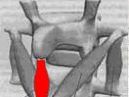
These muscles form a structure bordering the pubis and coccyx in front and behind, and on the sides with two ischial tuberosities. These muscles are arranged in the shape of a figure eight, in the center of which, at the intersection, is the central muscular point of the perineum, or the central tendon. The upper loop of the figure eight is located inside the front triangle.

bulbospongiosus muscle (bulbospongiosus) on the one hand, it is attached to the pubic bone and the shell of the clitoris, on the other hand, to the tendon center of the perineum, which is located between the vagina and the anus. This small muscle, about the size of a little finger, is considered the entry muscle because it closes the entry to the vagina when contracted. There is an assumption that this muscle has weakened as a result of the fact that it has lost its main meaning - to close the entrance of the vagina, in connection with such an achievement of civilization as the invention of underwear, which instead of this muscle began to protect the genital gap from dirt and dust.
When training this muscle during its contraction, you can feel the clitoris and even experience pleasant sensations.
It is believed that this muscle, being an entry muscle, during training creates the so-called “tight entry” effect. V.L. wrote about this in his books. Muranivsky. Another opinion is shared by Yu. Kornev and the International School of Imbuilding. According to imbuilding instructors, the "tight entrance" is created by the levator ani muscle due to the retraction of the anus.
muscles bulbospongiosus and ischiocavernosus cover cavernous bodies.
In the upper triangle there are two groups of muscles of the surface layer. They are located between two layers. perineal membrane.

The cavernous bodies are anchored below the deep layer of the membrane. These organs are made up of tissues rich in capillaries that can expand and swell. Cavernous bodies (corpus covernosum) are located along the branches of the pubic bones and are connected in front, forming body of the clitoris.
Clitoris- This is a cavernous body, similar to the male penis. It is located just behind the pubis and has the shape of a cylinder measuring 3 cm in length and 0.5 cm in diameter. (For more on the clitoris, see Pleasure Points.)
bulbs of vestibule located on both sides of the vulva.

bartholin glands located behind the back of the vulva. They highlight lubricant that flows into the vagina during intercourse.
DEEP LAYER OF THE PELVIC MUSCLES

These muscles are located deeper and higher than the previous level of the pelvic muscles, closer to the internal organs. This muscle group is shaped like an inverted dome. Its upper part is opposite diaphragm, this is where its name comes from. pelvic diaphragm.
pelvic diaphragm- this is, so to speak, the collective name of the muscles. These muscles form a kind of hammock, inside the domed surface of which all the pelvic organs are contained. They passively (elasticity) and actively (tonus) respond to various movements in the abdomen.
There are two muscles here:
- Muscle that lifts the anus (Levator ani)
- This is a strong muscle, consisting of various muscle bundles located around the openings of the internal organs. Its shape resembles a horseshoe.
This flat muscle has three bellies that sag like hammocks from the pubis to the sacrum. Sometimes this muscle is compared to a "tile roof", because. muscle belly overlap each other. Muscle fibers are woven into the rectum and braid the vagina from the sides and back. With its contraction, the anus rises, the vagina contracts, and the internal organs are also supported.
Muscle that lifts the anus, consists of two main parts.
- The middle part consists of puborectalis muscle (puborectalis)
, which starts at the pubis and wraps around the rectum, as well as pubococcygeus muscle (pubococcygeus)
, which runs from the muscular arch of the buttock and surrounds the rectum.
pubococcygeus muscle- this is also a "collective" muscle, which "entered" several muscles of the pelvic floor. Arnold Kegel united these several muscles into a single concept.
- The side part that bears the name iliococcygeal muscle (iliococcygeus)
, and consists of a large fibrous web. It runs along the posterior muscular arch and ends at the coccyx.
These knots of muscle tissue are of great importance for the maintenance of internal organs.

coccyx muscle coccygeus located on the same level with the muscle that lifts the anus. This muscle is stretched between the ischial spine, sacrum and coccyx.
 URINARY SINUS

In front, to the right and to the left of the insertion of the levator ani muscle, there is a recess, or notch, a zone in which there are no muscles. They call her urinary sinus. This zone corresponds to the junction of the bladder/uterus and the uterus/vagina.
Its physiological function is to serve as a wide passage for the fetus. But at the same time it is a weak link in the structure that supports the internal organs.
If the middle portion of the levator ani muscle (the puborectalis fasciculus) is well toned, it is able to actively support the bladder and uterus. Weakness of this zone almost always causes prolapse of organs and urinary incontinence (miction).
TWO LEVELS OF PELVIC FLOOR MUSCLES
The two layers of muscles mentioned above - superficial and deep - are located at different levels of the small pelvis.
They also differ in shape and direction.


The deep layer forms upper level. It is composed of muscles that form a broad surface attached to the median plane of the entry. As can be seen in the frontal cross section, this layer has the shape of a funnel, narrowed downwards.
The surface layer forms Lower level and consists of thin, woven muscular knots attached to the inferior plane of entry. As can be seen in the frontal cross section, this layer is located horizontally.
Located at the intersection of muscles central perineal tendon.
CENTRAL PERINETENDON

This is a zone of connective tissue, which includes most of the muscles in this area:
- deep transverse muscles of the perineum;
- bulbous spongy muscle;
- muscle that raises the anus;
- anal sphincter.
The central tendon is very elastic. During childbirth, this area is subjected to pressure, especially at the time when the fetal head erupts. It is in this place during childbirth, in case the fetus cannot come out, that a surgical procedure called episiotomy or, more simply, a cut.
You can read about the main female organs in 1 parts, about the structure of the bones of the female pelvis - in part 2 section "Lady's Anatomy". In the next part, we will talk about the main "Points of Pleasure".
The art of being a woman Frolova Evgenia Valentinovna
Learning the muscles of the pelvic floor
Learning the muscles of the pelvic floor
Consider how the muscular frame of the innermost female organs is arranged. The muscles of love in medical language are called the muscles of the pelvic floor. They are connected into a whole system, since in the body many internal muscles perform a vital function - supporting internal organs.
There are three diaphragms in our body. Above (approximately at the level of the solar plexus) is the respiratory diaphragm, which is responsible for inhaling and exhaling, rising and falling. Also, the urogenital and pelvic diaphragms. They work like hammocks that support all the abdominal organs. Just imagine for a moment what a weight it is - stomach, intestines, liver, kidneys, bladder! Therefore, everything in the abdominal cavity is supported by the pelvic floor muscles, which are so important for training that unlocks female potential.
Female reproductive organs
Muscles of the pelvic floor:
1. Bottom (outer) layer consists of muscles that converge in the tendon center of the perineum. In shape, they resemble a figure eight suspended from the bones of the pelvis. bulbospongiosus muscle ( m. bulbocavernosus) wraps around the entrance to the vagina, attaches to the tendon center and the clitoris. When this muscle contracts, the entrance to the vagina is compressed.
2. The middle layer is the urogenital diaphragm (diaphragma urogenitale), occupying the anterior half of the pelvis. It is a triangular muscular plate located in the pubic arch. The urethra and vagina pass through the urogenital diaphragm. In the anterior part of the urogenital diaphragm, muscle bundles surround the urethra and form its external sphincter. In turn, in the posterior section, muscle bundles are laid, running in the transverse direction to the ischial tuberosities. This part of the urogenital diaphragm is called the deep transverse perineal muscle ( m. transverse perinea profundus).
3. The anal muscles are the upper (inner) layer of the pelvic floor. , which is also called the pelvic diaphragm ( diaphragma pelvis). This layer consists of a paired muscle that lifts the anus ( m. levator ani). Both broad muscles that lift the anus form a kind of dome, the top of which is turned down and is attached to the lower rectum (slightly above the anus). The wide base of the dome is turned upwards and is attached to the inner surface of the walls of the pelvis. In the anterior part of the diaphragm, between the bundles of muscles that lift the anus, there is a longitudinally located gap through which the urethra and vagina exit the pelvic cavity ( Hiatus genitalis). The muscles that lift the anus consist of separate muscle bundles that start from different sections of the pelvic wall.
This layer of the pelvic muscles is the most powerful. If the anal muscles are in good tone, when emptying, a complete cleansing of both the rectum itself and the entire large intestine occurs.
With a weak work of these muscles, the intestines are not completely cleared, constipation begins, our body is poisoned by toxins. In this case, they begin to take laxatives, give enemas, and resort to hydrocolonotherapy. But the body can and should remove the decay products itself. And he does a great job with it if the anal muscles work as they should.
The development of hemorrhoids, that is, the bulging of the walls of the veins of the rectum, is also associated with detraining of the anal muscles. If they are in good condition, their tone is transferred to the walls of the colon and veins. Otherwise, the veins begin to bulge. (By the way, in the initial stages of hemorrhoids, this trouble can be dealt with by simply strengthening the anal muscles.)
The weakening of the pelvic floor muscles, which occurs due to their insufficient training or natural age-related changes, leads to a displacement of the pelvic organs. First of all, the functions of the bladder, uterus, fallopian tubes, ovaries, and sexual function suffer from this.
Almost all female gynecological problems are associated with the weakness of these muscles - stress urinary incontinence, prolapse of the uterus, vaginal walls, inflammation, tumor processes (uterine fibroids, endometriosis, etc.). How many people know that the correct location of the uterus depends on these muscles, without which the normal course of pregnancy and childbirth is almost impossible?
Infertility can also be associated with muscle weakness (often a long-awaited pregnancy occurs after their strengthening). During childbirth, all three layers of the pelvic floor muscles stretch and form a wide tube of the birth canal. If a woman wants to quickly restore her health after giving birth, then with the help of special exercises and simulators, her pelvic floor muscles quickly regain their original shape.
Insufficient tone of the pelvic floor muscles is the source of the vast majority of sexual problems: from general dissatisfaction with intimate relationships to the complete absence of orgasm.
As for men, their insufficient work of the muscular apparatus leads primarily to erectile dysfunction (from which impotence is within easy reach), as well as prostatitis and vasculitis. It will be quite good if men understand that trained pelvic floor muscles are:
Possibility of controlled ejaculation (increased duration of sexual intercourse); ? strengthening and strengthening erections, which increases your "rating" in women.
An erection occurs, of course, not only and not so much due to muscle tension, but as these muscles strengthen, its quality undoubtedly improves. In addition, when the muscles of the pelvic floor are tense, a natural massage of the prostate gland and seminal vesicles occurs. This reduces blood stasis in these organs and has an anti-inflammatory and resolving effect - something that was achieved earlier with the help of conventional manual prostate massage. So, we got a little acquainted with the muscles of the pelvic floor. If, while exploring ourselves, we dip our fingers into the vagina and try to contract the muscles that control the clitoris and side walls, then we will shrink 25% of the vagina. And if we now tighten the anus, then the remaining 75% will also be reduced.
From the book How to Get Married (From the first date to the wedding procession) author Kalinina OlgaSTUDYING THE OBJECT OF ATTENTION The more you know about men in general, the easier it is for you to understand a particular candidate. The more transparent their thoughts and behavior are, the more success you will achieve with the men you date. Know what their reaction to women
From the book Social Psychology. Intensive course. author Myers David JExploring Social Psychology Exploring Social Psychology David Myers is a psychologist of world renown. Myers' book is a masterpiece of teaching: in an engaging way, the reader is introduced to the science of human behavior in society, quickly and reliably.
From the book How to Influence People in Life and Business author Kozlov Dmitry AlexandrovichCHAPTER 2 STUDYING THE DISC INDIVIDUAL DIFFERENCE MODEL I didn't eat for days on end and didn't sleep all night long - I kept thinking, but in vain, it's more useful to study. CONFUCIUS So, let's go directly to the study of typology
From the book Enlightened Sex [Something Completely Different] author Deida Davidchapter thirty. closing the pelvic floor In its natural movement, energy flows from the genitals up the spine to the head and then down the chest and back to the pelvic floor. Both in sex and in everyday life, you can learn to "close the pelvic floor" (i.e.
From the book Autotraining author Alexandrov Artur AlexandrovichStress and Muscles Hans Selye noted that increased muscle contraction inevitably occurs under the influence of stress, and recommended using various methods of muscle relaxation to combat this contraction. However, he did not consider in detail the changes taking place in
From the book The Language of the Human Face author Lange FritzMuscles of the Nose The nose has some important muscles. They originate on the bones, which are located on the bone and cartilaginous plates and go directly into the skin of the nose (Fig. 36). Procerus originates in the middle of the back of the nose (Fig. 36, D), which is also called pyramidalis. At
From the book Maintaining Order in the Soul [A Practical Guide to Achieving Emotional Comfort] author Carrington-Smith SandraMuscles of the mouth The muscle that closes the mouth, orbicularis oris (final table, 4), forms the core of the muscular plate. Its fibers surround - like the fibers of orbicularis oculi - the labial fissure. When they contract, the mouth closes - the muscle is a sphincter. When the fibers are loosely tightened,
From the book The Power of Optimism. Why Positive People Live Longer author Clifton DonaldMuscles of laughter 1. M. risorius (final table, 9) The muscle starts below the zygomatic arch from the fascia that covers the parotis and masseter, stretching down to the corner of the mouth with a convex arch. It pulls the corner of the mouth outward and slightly upward, while simultaneously shifting the lower end of the nasolabial
From the book Mom and Baby. From birth to three years author Pankova Olga YurievnaFacial muscles Buccinator - trumpeter muscle, muscle of failure and disappointment, cheek muscle. Corrugator supercilii - muscle that wrinkles the eyebrows, tension muscle. Frontalis - frontal muscle, the second muscle of attention; medial bundle of fibers - pathetic pain muscle. Levator palpebrae superioris - lift
From the book Change Your Biological Age. Back to 25 author Lavrinenko Semyon ValerievichChapter 7 The Living Room Examining Our Relationships Almost all of our sorrows are caused by relationships with other people. Arthur Schopenhauer We successfully got rid of unnecessary trash in the house. What a huge achievement! The closets and the attic are organized, and we are no longer afraid that something necessary
From the book How? Earn on your image! Practical guide author Titov Dmitry YurievichExamining the Positive Shocked by such sophisticated cruelty and not wanting the unfortunate to be forgotten, Don Clifton and his colleagues set about studying the second part of the equation. They wondered: “If the constant negative impact is capable of completely
From the book Dudling for creative people [Learn to think differently] by Brown SunnyWe strengthen the muscles of the pelvic floor: work with a load To continue the exercises and increase the influx of sexual energy, I suggest working with a load. To do this, choose an egg of medium diameter, which has a special hole for the thread. Use as cargo
From the book I, again I and we by Little BrianRule number 1. We study the environment With age, a person is more and more susceptible to the "be like everyone else" syndrome. If youth is characterized by the desire to stand out from the crowd, then after 45 it’s the other way around, a person wants to be “no worse than others”. Maybe not worse. But not better, that's the thing. All in
From the author's bookStep 16 Learning how image formulas work Hey you! - the cat shouts to them on the run. - Now the carriage will pass here. If you do not say that these fields belong to the Marquis of Carabas, then you will not be in trouble. And don't try to sneak out. It will get worse! Charles Perrault "Puss in Boots" Part 1.
From the author's book From the author's bookWho is your city like? Exploring the Personalities of Cities and Regions In the previous pages, we have discussed comparatively objective characteristics of the environment, such as demographics, the number of incentives, and access to opportunities for social interaction. But places have
The pelvic floor is a powerful muscular-connective tissue plate and consists of three layers of muscles:
- the outer layer consists of 4 muscles (including the bulbous-cavernous muscle, which clasps and compresses the entrance to the vagina during contraction, and the sphincter of the anus - a circular muscle that "locks" the rectum);
- the middle layer is the urogenital diaphragm. The urethra and vagina pass through it. Contains the sphincter of the urethra - a circular muscle that "locks" the urethra;
- the inner layer consists of muscles that lift the anus. With their contraction, the genital gap closes, the lumen of the vagina and rectum narrows.
What is pelvic floor rehabilitation?
Pelvic floor rehabilitation is a set of activities aimed at strengthening the muscles of the pelvis.
Why does pelvic floor muscle weakness occur?
After vaginal delivery, the vagina usually expands somewhat, and its elasticity decreases to some extent. Childbirth, especially complicated ones, lead to damage (stretching, tears, ruptures) of the pelvic floor muscles. When the perineum is ruptured or dissected (episio- or perineotomy), the muscles of the inner layer are especially often damaged, sometimes after restoring the integrity of the perineum, the genital gap does not completely close. At the same time, with age, the muscles of the pelvis, as well as the muscles of the whole body, weaken.
What causes weakness in the pelvic floor muscles?
Weakening of the pelvic floor muscles, as well as impaired contractility of these muscles, leads to conditions such as urinary incontinence, prolapse of the anterior and posterior walls of the vagina, prolapse of the uterus, chronic pelvic pain, soreness in the vestibule of the vagina. A decrease in the elasticity of the tissues of the vagina and a decrease in the sensitivity of the tissues of the perineum can lead to a decrease in sexual sensations in both partners.
What is urinary incontinence?
Urinary incontinence is the involuntary loss of urine.
How common is urinary incontinence in the world?
About 40% of women over 40 suffer from urinary incontinence, and only 4% do not consider this phenomenon to be natural.
What are the types of urinary incontinence?
According to the International Continence Society, there are six types of urinary incontinence:
1. Stress urinary incontinence (stress incontinence) - involuntary release of urine during physical exertion, coughing, sneezing, etc., i.e. in cases of a sharp increase in intra-abdominal pressure.
2. Urgent urinary incontinence - involuntary release of urine with a sudden, strong and unbearable urge to urinate.
3.Mixed urinary incontinence - a condition that combines the symptoms of the first two types of urinary incontinence.
4. Nocturnal urinary incontinence (enuresis).
5. Involuntary leakage of urine, not accompanied by an urge to urinate.
6. Other situational types of urinary incontinence (for example: urinary incontinence during sexual intercourse, when laughing, etc.).
What is the normal mechanism for continence of urine?
Normal retention of urine is carried out through the interaction of four main mechanisms:
1. stable position in the body of the bladder;
2. immobility of the urethra;
3. adequate innervation of the pelvic floor muscles and the muscular membrane of the bladder;
4. anatomical and functional integrity of the closure apparatus of the bladder and urethra.
What are the risk factors for urinary incontinence?
Pregnancy, childbirth.
Gender - More common in females.
Age - more common after 40 years.
Increased weight.
The hereditary factor is a genetic predisposition to the development of urinary incontinence (connective tissue dysplasia syndrome).
Neurological factor - the presence of various diseases of the nervous system.
Anatomical factor - anatomical disorders of the pelvic floor muscles and pelvic organs.
Surgical interventions - damage to the pelvic nerves or muscles.
What is the most common type of urinary incontinence?
The most common type of urinary incontinence is stress urinary incontinence - the involuntary release of urine during physical exertion, coughing, sneezing, etc., i.e. in cases of a sharp increase in intra-abdominal pressure. In this case, urinary incontinence is usually combined with a weakening of the pelvic floor muscles, so the treatment of stress urinary incontinence must be combined with therapy aimed at the rehabilitation of the pelvic floor muscles.
What are Kegel exercises?
The exercises proposed by Arnold Kegel are aimed at training the muscles of the pelvic floor. These exercises can be performed independently, without the presence of a doctor. They do not require special clothing or equipment. They can be performed almost anytime and anywhere convenient.
How to do Kegel exercises?
To understand which muscles need to be reduced, you need:
Try to interrupt the stream of urine while urinating.
Contract the same muscles as if to interrupt urination, but do this outside of urination.
Squeeze the muscles of the rectum as if to prevent the release of gases. However, the buttocks must remain motionless.
Important: when performing exercises, you can not connect the abdominal muscles. Legs and buttocks should remain motionless.

Kegel exercises:
1. Strongly contract the vaginal muscles for 1-2 seconds, then relax them; to achieve the effect, it is necessary to perform several times a day for 5-30 contractions.
2. Contract the vaginal muscles for 10 seconds, then relax for 10 seconds. Do the exercise 4 minutes a day. After that, make quick contractions for 1 minute (1 second each), alternating them with the same quick relaxations.
3. Exercise "lift": contract the muscles of the vagina ("1st floor"), hold for 3-5 seconds, continue contraction with greater force ("2nd floor"), hold again. So go 4-5 "floors". Perform the same step-by-step movement "down", lingering on each "floor". You can perform exercises at home, in transport, while watching TV.
How to do Kegel exercises correctly?
Do these exercises as often as possible. The more often, the better the result will be.
Start doing exercises with a five-second interval, holding the muscles in a contracted state for five seconds. Gradually lengthen the contraction time.
Important: do not stop exercising, despite muscle fatigue.
When can exercise results be evaluated?
Immediate improvement can be felt within a few weeks of starting exercise. However, to obtain a reliable result, it is necessary to perform the exercises for at least 4 months.
What to do if the exercises did not work?
If gymnastic exercises have not brought positive results due to poor muscle sensation, training with vaginal cones can bring the desired result.
What are vaginal cones?
Vaginal cones are specially designed conical weights with variable weights that are used to strengthen the muscles of the vagina. The cone is placed in the vagina like a tampon. A set of four cones with different weights has been developed. The woman's task is to learn how to hold the cone by contracting the muscles of the pelvic floor.
To prevent prolapse of the walls of the vagina and the body of the uterus, as well as other pelvic organs;
- during pregnancy and after childbirth to prevent weakening of the pelvic floor muscles and restore their original tone;
- in order to reduce the risk of infection and the ingress of unfavorable flora, as well as changes in the pH environment of the vagina with a gaping genital slit;
- to eliminate and prevent urinary incontinence (stress urinary incontinence when coughing, sneezing, physical activity);
- to control the force of contraction and relaxation of the vaginal muscle group in order to increase sexual sensations during intercourse. Long-term training with vaginal cones promotes the sensation of the pelvic floor muscles and their development. During pregnancy, cone training develops the pelvic floor muscles and prevents them from weakening.

How to use vaginal cones?
It is necessary to choose a cone of the appropriate weight (for starters - the lightest). Insert it into the vagina with your index finger, in the same way as a vaginal tampon.
It is necessary to keep the cone in a standing position.
If it works, then:
Hold the cone by taking a few steps.
If it works, then:
Hold the cone while walking.
It is necessary to fix how much time it is possible to hold the weight. The holding time should be gradually increased.
Hold the cone while walking up the stairs.
Hold the cone while coughing.
Hold the cone throughout the day.
How often and for how long should you do exercises with vaginal cones?
These exercises should be done at least twice a day, and, if possible, more often. If the cone is held quietly during the day, you can increase the mass of the vaginal cone. Thus, it is necessary to achieve the ability to hold the heaviest cone without straining during normal daily activities.
Can I do Kegel exercises with vaginal cones?
The combination of Kegel exercises with the use of vaginal weight cones is very effective.
- It is possible to perform contractions of the pelvic floor muscles with a cone placed in the vagina.
- It is necessary to move to the next weight if there is an ability to perform the exercise for at least 5 minutes.
- You can lengthen the exercises up to 10 minutes, going back to the lightest weight, and so on.
What to do if it is impossible to keep the vaginal cone in a standing position?
Start doing exercises lying down. In the future, having strengthened the muscles of the pelvic floor, you can move on to a standing position.
What are the benefits of using vaginal cones:
- Individuality for every woman.
- It takes little time to teach a woman how to use cones.
- It takes little time to start training.
- The number of consultations with a doctor is reduced to one visit.
- Cones are one form of biofeedback.
- The weight of the cones can be increased by increasing the load.
- You can start using it without additional research.
How to evaluate the effectiveness of the use of cones?
If gymnastics for the muscles of the pelvic floor is carried out under the supervision of a doctor, with urinary incontinence or in the presence of gynecological problems, then the doctor can evaluate the effectiveness of the treatment both according to the results obtained (symptoms will disappear) and with the help of special digital devices - perineometers. The vaginal sensor is inserted into the woman's vagina, then she contracts the muscles of the perineum as much as possible, and a quantitative assessment of this contraction is displayed on the scale of the device. The perineometer works on the same principle as a blood pressure monitor, meaning it measures the pressure generated in the vagina.
How long should the cones continue to be used?
With regular daily use, improvement is noticeable after 8 weeks. To get the most out of your cones, it's important to train every day for a minimum of 12 weeks.
Can cones be used for vaginal dryness?
A small amount of lubricant must be applied.
What to do after the pelvic floor muscles have become stronger?
How soon after having a baby can I start using Kegel exercises and cones?
Kegel exercises and cones can be started as soon as the woman feels comfortable after childbirth. On average, training is recommended 6-8 weeks after birth.
- It is necessary to rinse the cone before each use (to prevent irritation or infection).
- It is best to insert the cone after emptying the bladder.
- It is necessary to wear underwear: if the cone falls out, it will not be lost.
- If possible, practice with the cone at the same time of the day. It is convenient to perform exercises at the same time as your usual daily activities.
- If it is impossible to hold the lightest weight, place your finger on the tip of the cone (where the thread is attached to it), this will reduce the weight. Then do the exercises.
- If it is possible to keep the cone inside the body for 15 minutes, you can try to go up and down the stairs, do household chores. Such actions can really teach you to control the function of the bladder. You may need to use lighter cones when performing these steps.
- Always remove the cone after use. It is intended to be used for limited periods of time, during the daytime, and should not be used continuously.
Are there any contraindications for using cones?
The cones are not intended for use by women suffering from or suspected of having a disease in the vaginal, genital or pelvic area (infections, inflammatory diseases, malignancies of the pelvic organs). Cones should not be used during the first six weeks after childbirth or pelvic surgery. It is not recommended to use cones during or immediately after sexual intercourse, as well as during menstruation. Do not use cones at the same time as tampons, uterine ring or diaphragm.
What to do if there is no effect?
Can exercises and cones be used to prevent pelvic floor muscle weakness?
Exercise is useful for almost all women, not only those with signs of weakness of the corresponding muscles. Their implementation is the prevention of chronic inflammatory diseases of the small pelvis, venous stasis, prolapse of the walls of the vagina, urinary incontinence, increases sensitivity during sexual activity. In addition to the prevention of many gynecological diseases, they also help prevent the weakness of labor activity (training of the intimate muscles is desirable before childbirth, during pregnancy, given the load ahead in childbirth), the initial stages of urinary incontinence.
What are the treatments for stress urinary incontinence?
Conservative - special exercises to strengthen the muscles of the pelvic floor, discussed above.
The goal of the surgical treatment of stress urinary incontinence is to create additional support for the urethra in order to eliminate the pathological mobility of the latter. The choice of one or another method largely depends on the degree of urinary incontinence.
What is a loop (sling) operation?
There are many options for loop (sling) operations, during which the effect of urinary retention is achieved by creating a reliable additional support for the urethra by placing loops of various materials under the middle part of the urethra (vaginal flap, skin, cadaveric fascia, etc.). Recently, minimally invasive loop operations have become increasingly popular, which have certain advantages (TVT operation, TVT-O operation, TOT operation, etc.).What are the benefits of minimally invasive surgeries?
- Good tolerability - used for any degree of urinary incontinence.
- Use of a synthetic mesh as a loop material.
- Opportunity to perform surgery under local anesthesia.
- The short duration of the operation (about 30 - 40 minutes).
- Short postoperative period - the patient can be discharged home on the day of surgery or the day after surgery.
- Good functional results - low probability of recurrence of the disease.
What is pelvic floor reconstruction?
Pelvic floor reconstruction is a surgical operation aimed at correcting defects in the pelvic floor that cannot be treated conservatively. Pelvic floor reconstruction remains one of the most difficult problems that brings together the efforts of urologists, gynecologists and proctologists.What is the purpose of pelvic floor reconstruction surgery?
Operations for reconstruction of the pelvic floor allow you to restore the normal anatomical relationships of the pelvic organs with the help of the woman's own tissues or special synthetic materials that strengthen the pelvic floor. These operations are used for prolapse of the bladder, uterus, vaginal vaults, and other types of violations of the anatomy of the pelvic floor. In most cases, these operations avoid the removal of the uterus when it is significantly lowered. Vaginoplasty is performed under general anesthesia, spinal or epidural anesthesia. The average duration of the operation is 1-1.5 hours. During the intervention, the patient does not feel pain.
What happens to the implant in the future after mesh plasty?
Prolene mesh does not dissolve and does not break down under the action of enzymes and retains its strength and integrity throughout the patient's life. Being essentially inert, the mesh causes the formation of a thin layer of fibrous tissue that can grow through the pores of the mesh. Germination of fibrous tissue leads to a stronger connection of the mesh with surrounding tissues. Large pore sizes do not prevent the migration of macrophages to the area of inflammation, thus preventing infectious complications.
What are the features of the postoperative period after reconstructive surgery on the pelvic floor?
- The toilet of the external genitalia and perineum is performed 4-5 times a day;
- vaginal douching is not recommended;
- stitches on the skin of the perineum are removed on the 5th day;
- a woman is recommended to eat easily digestible food to prevent constipation ("straining" after surgery is contraindicated);
- sitting is allowed only 15-20 days after discharge from the hospital. It is not allowed to lift weights (more than 5 kg) and physical activity should be limited.
Sex life is allowed 2 months after the operation.
Pregnancy should not be planned within the next 12 months after surgery. During subsequent births, vaginal ruptures along the old scar are not excluded, but this is rare, since the tissues have time to restore their anatomical and functional usefulness. The presence of vaginal plasty is not an indication for a caesarean section in the future.
When should you not have surgery?
- For all general conditions of the body in which planned surgical interventions are contraindicated (fever, infectious diseases, oncological pathology, some blood diseases);
- in the presence of a mild degree of identified violations, which makes it possible to successfully apply conservative methods of treatment.
Symptoms that should see a specialist:
- violation of the physiological functions of the pelvic organs of varying degrees (bladder, rectum), which can appear both in the postpartum period and during pregnancy - incontinence of urine, gases, feces;
- lack of previous sensations during sexual activity in the presence of desire (anorgasmia), painful sensations during sexual intercourse;
- gaping of the genital slit, sometimes causing dryness in the genital area;
- the appearance of complaints in connection with a violation of the microflora of the vagina, the urethra (periodically increasing mucous whitish discharge with an unpleasant odor in the absence of any urinary tract infections);
- prolapse of the walls of the vagina and uterus (usually detected during a gynecological examination)
perineum, or pelvic floor, called the muscular-fascial plate that closes the exit from the small pelvis, its lower aperture.
The rhombus-shaped perineum is bounded in front by the pubic symphysis, from the sides by the ischial tuberosities, and from behind by the coccyx. The line connecting the ischial tuberosities divides the perineum into two triangular shaped areas.
The anterior region is called urogenital diaphragm, and back - pelvic diaphragm. The urethra passes through the urogenital diaphragm in men and the urethra and vagina in women. The end section of the rectum passes through the pelvic diaphragm.
Both the urogenital diaphragm and the pelvic diaphragm are formed by two layers of muscles and the fascia that covers them.
At pelvic diaphragm in the surface layer, there is an annular unpaired muscle - external sphincter of the anus, voluntary muscle that closes the exit from the rectum. In the second, deep layer, there is a triangular steam room muscle that lifts the anus. The muscle begins on the inner surface of the walls of the pelvis, goes down and is woven into the final section of the rectum. Both muscles that lift the anus surround the lower part of the rectum in the form of a funnel.
urogenital diaphragm also has superficial and deep layers of muscles and fascia. In the surface layer are paired superficial transverse perineal muscle, as well as bulbous-spongy and sciatic-cavefrizzy muscles, that promote erection of the penis or clitoris. The bulbospongiosus muscle in men surrounds the bulb and spongy body of the penis, and in women it surrounds the entrance to the vagina. Ischiocavernosusmuscle, starting on the ischial tuberosity, is woven into the cavernous body of the penis in men or the clitoris in women. In the deep layer of the urogenital diaphragm there is an unpaired muscle - sphincter (external) urinationboiler channel and steam room deep transverse muscleboundaries.
The muscles of the perineum are covered on the outside superficial fascia of the perineum. In addition, the muscles that lift the anus and its sphincter are enclosed between bottom and superior fasciae of the pelvic diaphragm. The deep transverse muscles of the perineum and the sphincter of the urethra are covered bottom and upper fascia of the genitourinarydiaphragm.
Muscles and fascia of the head
The muscles of the head, taking into account their location and functions, are divided into two groups: facial muscles and chewingbody muscles.
Mimic muscles
They are located under the skin and are grouped in the form of constrictors and dilators around the mouth and nasal openings, eye sockets, external auditory canal. The mimic muscles do not have superficial fascia. Mimic muscles begin on the bones of the skull and are woven into the connective tissue base of the skin. With their contraction, the facial muscles shift the skin, forming dimples, furrows, folds. Thus, the mimic muscles form complex expressive facial movements - mimiku. According to the location, the facial muscles are divided into:
supracranial muscle has two abdomens - frontal and occipital, between them is an extensive supracranial aponeurosis.
- occipital abdomen begins at the highest line of the occipital bone, it pulls the entire muscle backwards, smoothes the wrinkles on the forehead.
- frontal abdomen is woven into the skin of the forehead, when it is reduced, the eyebrows rise and transverse folds of the skin of the forehead are formed - wonder muscle,interrogative attention.
Eyebrow wrinkling muscle located in the thickness of the eyebrow, begins on the nasal part of the frontal bone, is woven into the skin of the eyebrow. With bilateral contraction, these muscles bring the eyebrows together, form facial expressions of pain, suffering, gloom.
Muscle of the proud located on the bridge of the nose, when reduced, it forms a transverse fold over the bridge of the nose.
Circular muscle of the eye formed by circular bundles surrounding the orbit - eye part, weaving into the skin of the eyelids - eyelidthe main part and attached to the lacrimal sac - tearspart. With its contraction, the muscle closes the eyelids, closes the eyes, promotes the outflow of tears into the nasolacrimal canal.
Muscles surrounding the nasal passages begin on the anterior surface of the upper jaw, are woven into the wing and the cartilaginous part of the back of the nose. These muscles constrict the nostrils.
The muscles surrounding the opening of the mouth are formed by dilators and bundles located in the thickness of the lips - constrictors. Some dilators are located above the oral fissure - muscles that lift the corner of the mouth, muscles that raise the toplip, zygomatic muscles (small and large), muscles of laughter. These muscles form facial expressions of a smile, laughter. Other muscles lie below the oral fissure - muscles that lower the corner of the mouth, muscles that lower the lower lip. Here is located chin muscle that forms a hole.
buccal muscle, forming the muscular basis of the cheek, begins on the back of the upper and lower jaws, is also woven into circular muscle of the mouth. This muscle is involved in the act of sucking, moving the food bolus to the pharynx, straining the cheek, in connection with which it was called muscles trumpeter.
Mimic muscles include rudimentary mouseauricle-anterior, superior and posterior earmuscles, well developed in animals, wide flat subcutaneous muscle of the neck and others.